Warm weather has arrived, and many people will be spending more time outdoors in the sun. That can be good and bad.
“Even one blistering sunburn during childhood or adolescence can nearly double a person’s chance of developing melanoma,” the American Academy of Dermatologists website reads. “Experiencing five or more blistering sunburns between ages 15 and 20 increases one’s melanoma risk by 80 percent and nonmelanoma skin cancer risk by 68 percent.”
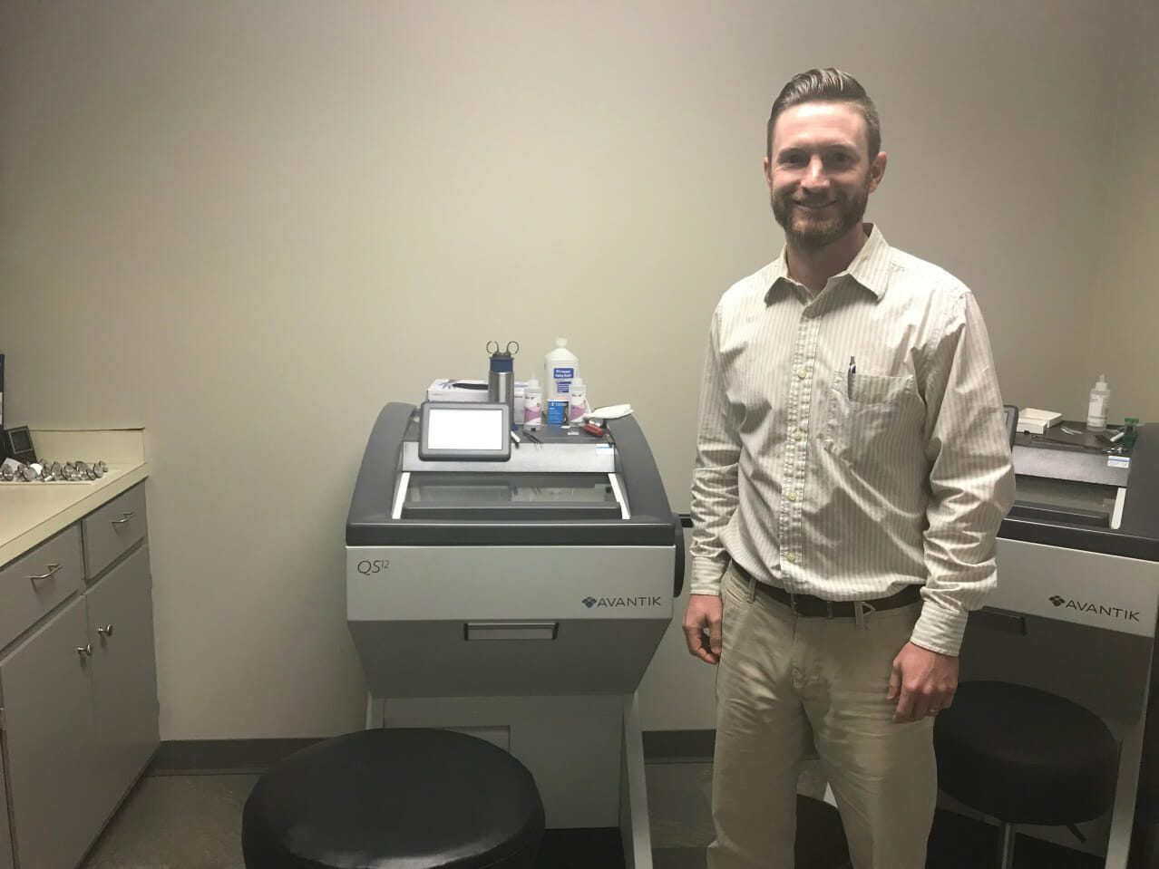
Dr. Brad Greenhaw and nurses, Ashley Graves and Jenna Cook, at Tupelo’s Dermatology Center see patients facing many skin/hair issues, and they spend most days using the cryostat.
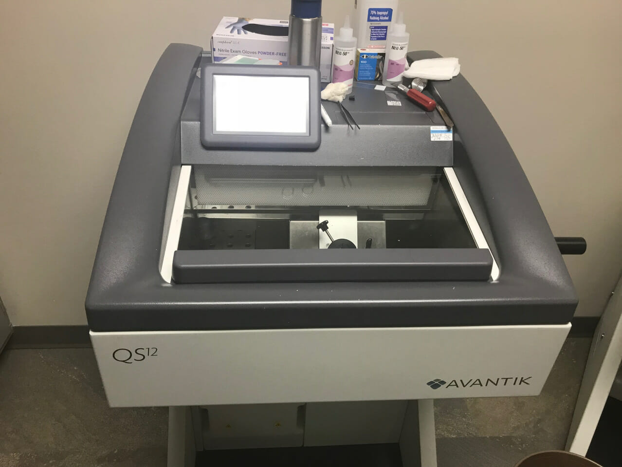
The cryostat is a machine used to cut tissues into thin slices while keeping it cold. The tissue is then viewed under a microscope so doctors and nurses can identify and treat skin cancer. The cryostat is an ultrafine tissue slicer placed in a freezer.
Greenhaw said he enjoys working in the dermatology field. “It’s a field where we, as doctors, can cure cancer,” he said. “I like the challenge of it, and there’s a lot of variety in what I do.”
According to the American Academy of Dermatology website, skin cancer is the most common cancer in the United States. It is estimated that one in five Americans will develop skin cancer in their lifetime.
Melanoma rates in the United States doubled from 1982 to 2011, the AAD website reports. Basal cell and squamous cell carcinomas, the two most common forms of skin cancer, are highly curable if detected early and treated properly. Most skin cancer deaths are from melanoma.
The AAD reports that approximately 95 percent of melanoma cases are attributable to UV exposure. In 2010, new research found that daily sunscreen use cut the incidents of melanoma, the deadliest form of skin cancer, in half.
The cryostat helps in the diagnosis and treatment of skin cancer. Instead of sending tissue cells to pathology labs and having pathologists cut them into little slivers in which the whole tissue can’t be clearly seen, Greenhaw said the cryostat cuts tissue on its side so tissue is visible from a thinly-sliced side view.

“We have a clearer and actual whole view inside of the tissue compared to what pathologists might see,” said Greenhaw. “They cut a little sliver and could miss tissue that can’t even be seen. Whereas here, we cut the side and can get a full perspective … The difference is how the tissue is processed and looked at through the scope.”
Unless doctors have their own cryostat to use at their convenience, which Greenhaw said most dermatologists do not, it is sent to the pathology lab. “Every day we do surgery and use it,” Greenhaw said. “It’s usually two days a week, but mostly three days a week.”
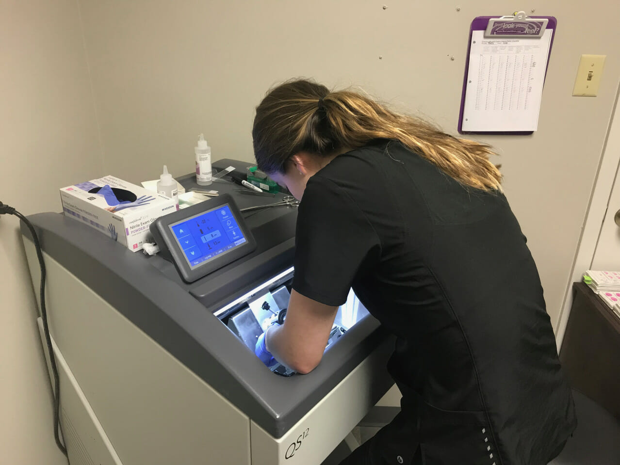
Nurses Cook and Graves operate the machine. “We bring (the tissue sample) in on a white tray, put it on a slide, and put it into the cryostat,” Graves said. “Once that is done, we then freeze the slide down to a chuck or block. After it’s frozen down, we then put the slide in the center of the machine and slice down the tissues.”
They write the patient’s name on the slide. “We do four slides depending on the size of the piece,” Graves said. “We usually do seven to eight more case surgeries in a couple of hours just to get the first stage.”
Greenshaw examines them. Nurses cut the tissue into sections, mount it on the slides, and put them on a burner for four minutes. They repair the slides in the order they are received.
“You keep going or repeating the process until the cancer is cleared out,” Graves said. “The slides are then put in a stainer after they burn, and that is how the cancer cells are identified.”

Stains that are used in the staining process include eosin, bluing, hydrochloric acid, hematoxylin, methanol, water and 100 percent reagent alcohol.
“You never know how long it is going to take,” Graves said. “We keep two cryostats going at the same time. It really depends on the volume of the tissue and how many patients Dr. Greenhaw sees that have to have the procedure done. We also do melanomas, but we mostly focus on basal cells or squamous cells.”
Graves said there is a different staining process for doing basal and squamous cells. “We cut pieces with fat, with muscle, some glands, and some with nerves,” she said. “It’s the majority of what we do here at the dermatology center. I love it. It’s pretty interesting, but it can be tedious.”
Overall, Dr. Greenhaw and his nurses hope to treat skin cancers and ensure their complete removal.
By Maggie Houin
Read more stories like this on Oxford Stories.
For questions or comments, email hottytoddynews@gmail.com.

